Introduced medical equipment
- Robot assisted surgery“da Vinci surgical system”
- URONAV(MRI/US fusion biopsy)
- Photometric diagnostic system for superficial bladder cancer
- Extracorporeal shock wave lithotomy (ESWL) device
- Laparoscopic surgery system
- Ingenuity Elite 64-row 128-slice CT scanner
- Roentgen equipment
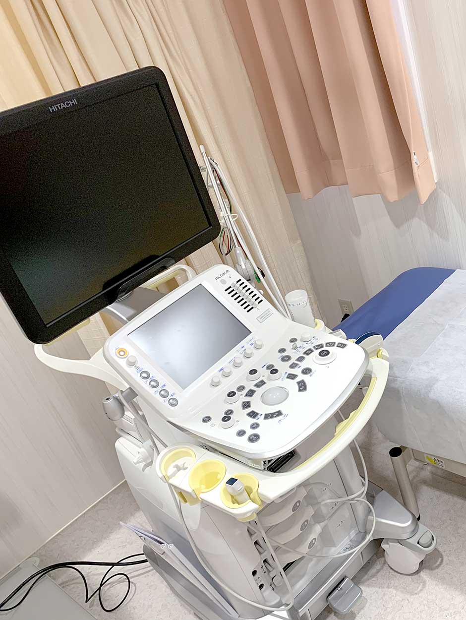
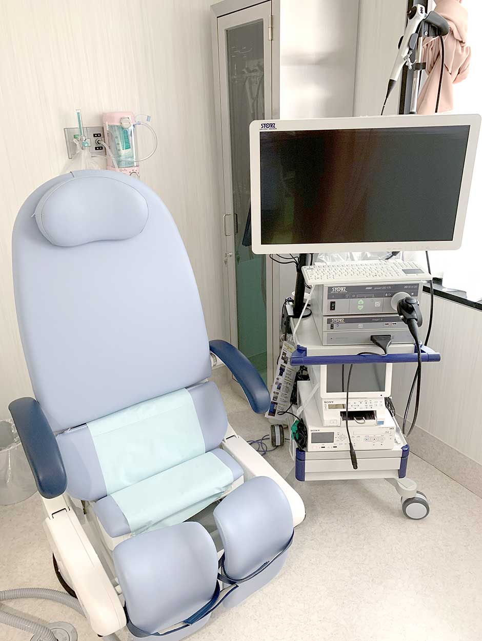
- Other inspection equipment
Robot assisted surgery“da Vinci surgical system”

Robotic surgery allows doctors to perform many types of complex procedures with more precision, flexibility and control than is possible with conventional techniques. Robotic surgery is usually associated with minimally invasive surgery — procedures performed through tiny incisions. Robotic surgery has been rapidly adopted by hospitals in the Japan for use in the treatment of a wide range of conditions.
Robot surgery centerURONAV(MRI/US fusion biopsy)


At our hospital, we have introduced an “MRI-Ultrasound fusion image” system (“UroNav”) which is one of the four facilities in Japan.
UroNav” has the function of fusing an MRI image with ultrasound images in real time and displaying it as a 3D image, and transrectally ultrasound the site where cancer is suspected by MRI. It is now possible to perform anterior gland examination while displaying on the sound image and directly confirming the inspection site on the super-wave image (MRI¬ super-wave elastic fusion: elastic fusion).
As a result, we succeeded in greatly improving the positive predictive value of cancer to 80% or more. * Positive predictive value is a positive test rate of 80% or more when MRI is positive (PIRADS score 4 points or more).
Photometric diagnostic system for superficial bladder cancer
When performing bladder cancer surgery, small cancers and flat cancers were difficult to confirm with conventional endoscopes. After the patients have a 5-aminolevulinic acid at 2 hours before surgery, cancer is fluoresced brightly by using a cystoscope. This technique leads the diagnosis for cancer more reliably Tokyo International Ohori Hospital has introduced this technology and aims to more reliably remove cancer and reduce recurrence as much as possible by using photometric diagnosis during TUR-BT. (Not all patients with superficial bladder cancer use this technique).
When making this photometric diagnosis, it is necessary to avoid a direct sunlight for two days after surgery in order to eliminate side effects (photosensitivity, etc.).


The prognosis of superficial bladder cancer varies depending on the malignancy and progression of the cancer, but when treated with transurethral resection, a cure rate of about 80% or more is obtained.
However, the recurrence rate is said to be 20 to 30% even if the patient is cured by treatment such as intravesical injection therapy.
Therefore, it is very important to follow up regularly with urinalysis, urine cytology, cystoscopy, etc. to detect recurrence at an early stage.
These tests are usually done every 3 months for the first 2 years, every 6 months for the 3rd year, and every year thereafter.
If recurrence is observed, transurethral resection will be performed again.
Extracorporeal shock wave lithotomy (ESWL) device

ESWL is a device that applies shock wave energy to the water from outside the body and crushes it into stone. At our hospital, we have introduced the EDAP TMS ESWL “Snolith I -move”. It is possible to identify a small stone ao that early treatment (crushing stone) is possible.


Laparoscopic surgery system

In addition to robot surgery, we also perform laparoscopic surgery. Since it is a minimally invasive procedure that does not burden the patient’s body, we may guide you through laparoscopic surgery depending on the case of the procedure. We always perform procedures that are meaningful to patients and have no disadvantages.
Ingenuity Elite 64-row 128-slice CT scanner

The CT scanner introduced at this hospital is Philip’s highest-end model that has both high image quality, speed, and appropriate low dose. Since it is possible to perform speedy examinations compared to conventional CT scanners, even patients who have difficulty in respiratory arrest can easily perform examinations. In addition, it is possible to collect detailed data in detail, which is effective for early detection of diseases that were difficult to detect until now.
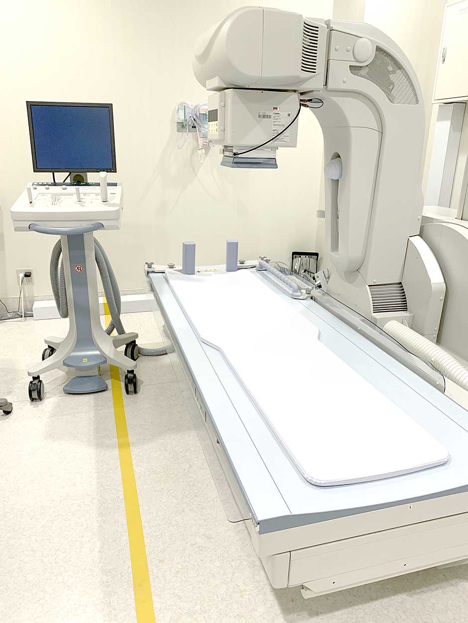
Roentgen equipment


Other inspection equipment